
Photo-mediated ultrasound therapy (PUT)
Introduction:
Angiogenesis plays a critical role in the pathogenesis of numerous pathological conditions, including cancer, inflammation, and eye diseases. Currently available anti-vascular treatments such as anti-VEGF agents and photodynamic therapy have several drawbacks. We have developed a novel, selective, localized anti-vascular technique, namely photo-mediated ultrasound therapy (PUT), by applying synchronized nanosecond laser irradiation and ultrasound pulses. Taking advantage of the high optical absorption of hemoglobin, PUT can selectively target microvessels without causing unwanted damages to the surrounding tissue. Moreover, PUT is totally noninvasive and agent-free.
The mechanism of PUT is thought to be cavitation activities in microvessels. Cavitation is produced via photospallation due to transient thermoelastic stress following short pulse duration laser and is driven by concurrent ultrasound. The addition of a laser pulse to an existing ultrasound field can significantly improve the likelihood of localized inertial cavitation, which can induce micro-vessel damage through its mechanical effect. In PUT, the required energy levels of both ultrasound and laser are significant lower than those used in previous therapeutic effects. Therefore, no significant temperature rise is induced and thermal damage can be effectively avoided.
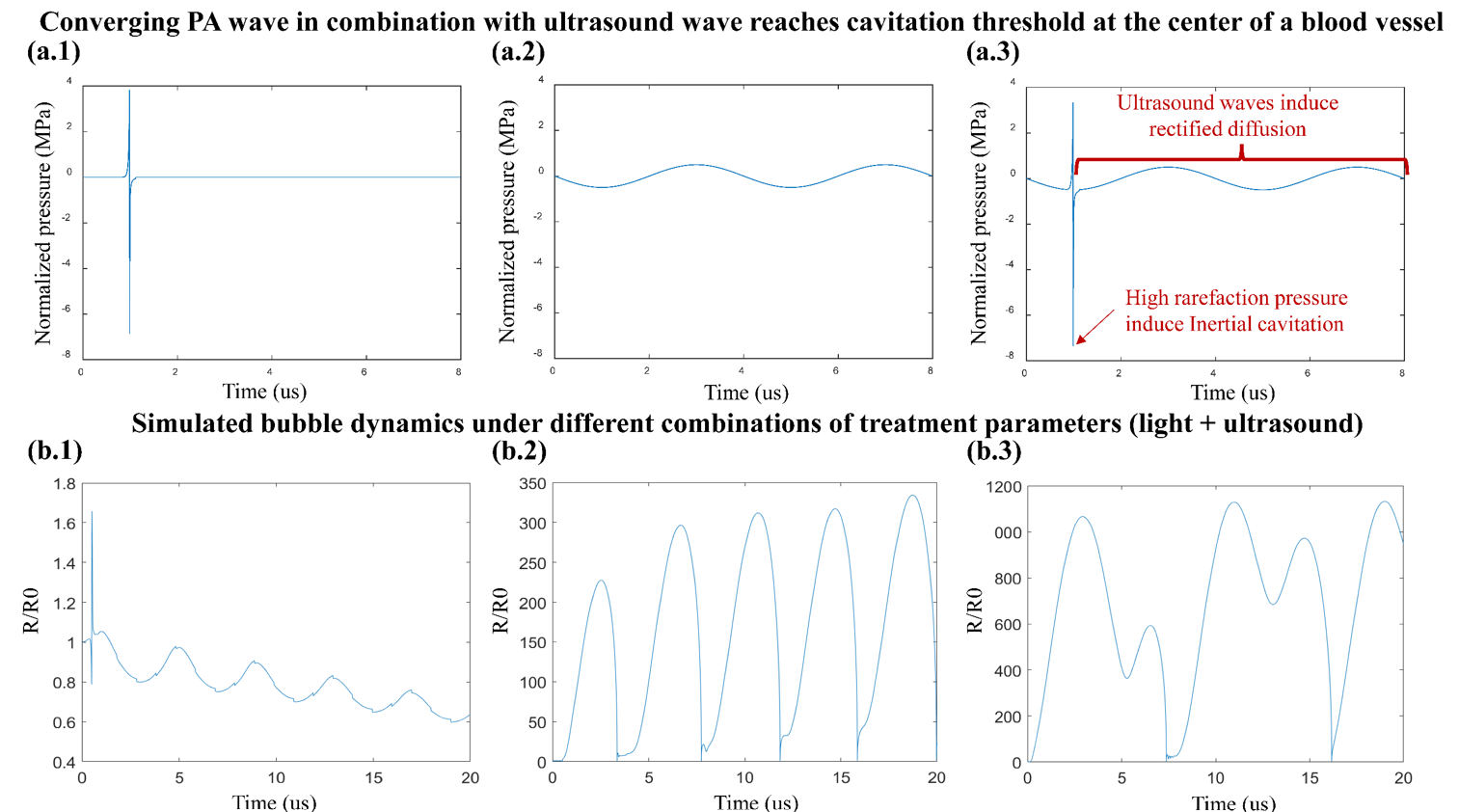
Figure 1. Theoretical modeling of the pre-existing bubble dynamics under different light fluence and ultrasound pressure during PUT treatment of a blood vessel. (a.1) Simulated photoacoustic (PA) wave near the center of a blood vessel with a diameter of 0.1 mm when illuminated by a 3-ns light pulse at 1064 nm wavelength. (a.2) Concurrently applied ultrasound burst with 0.25 MHz frequency and 1 MPa pressure amplitude. (a.3) Combined PA waveform in (a.1) and ultrasound wave in (a.2), where the PA wave is synchronized at the negative phase of an ultrasound cycle. (b.1-b.3) Simulated bubble size evolution under different combinations of treatment parameters, including 0.6 MPa ultrasound and 100 mJ/cm2 light fluence (b.1), 0.8 MPa ultrasound and 120 mJ/cm2 light fluence (b.2), and 1.0 MPa ultrasound and 120 mJ/cm2 light fluence (b.3). R/R0 stands for dynamic bubble size R over its initial size R0.
We are developing PUT as a novel method to selectively treat pathologic microvasculature in retinal vascular diseases, in addition to investigating other applications in tumors and dermatologic applications, including port wine stains and tattoo removal.
Example results:
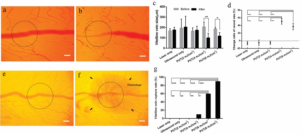
TFigure 2 The vein on a chicken yolk sac membrane before treatment with PUT (a). The vein after being treated by PUT with 0.45 MPa ultrasound and 4 mJ/cm2 laser fluence at 532 nm (b). The black circle corresponds to the region of treatment. Change in blood vessel diameter after laser only, ultrasound only, and PUT at 2, 4 and 6 mJ/cm2 fluence (c). Vertical bars demonstrate standard deviation. The relative change of the vessel size shows that statistical significances exist between 4 and 6 mJ/cm2 PUT and any other group (d). Higher laser fluence can result in vessel rupture (e,f,g). Repeated 10 times for each group. *p < 0.05; **p < 0.01, ***p < 0.001. Scale bar: 200 µm. Courtesy of the Authors.
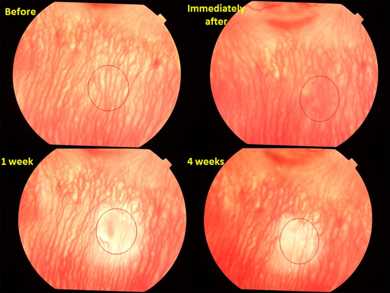
Figure 3 Fundus photos of rabbit eye before and after PUT treatment. The rabbit choroidal vasculature is treated by PUT with 2 MPa ultrasound + 75 mJ/cm2 laser, which caused edema immediately after treatment. By 1 week, pallor occurred in the region of treatment with greatly diminished choroidal vessels which persisted to 4 weeks.
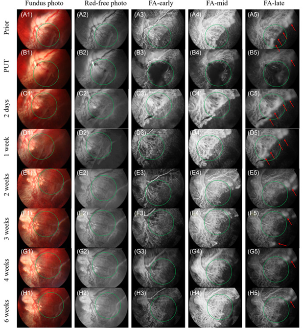
Figure 4. Representative fundus photographs (A1–H1), red‐free images (A2–H2), and fluorescein angiography (FA) of DBP rabbits’ RNV with single PUT treatment. (A3–H3): Early phase FA. (A4–H4): Middle phase FA. (A5–H5): Late phase FA. In these images, the extensive leakage (red arrow) indicated the region of RNV. Prior: 1 day before the PUT treatment (8 weeks after intravitreal DL‐AAA injection). PUT: immediately after the PUT treatment. Two days: 2 days after PUT treatment. * weeks: * weeks after PUT treatment (* = 1, 2, 3, 4, 6). The green circles indicate the PUT treatment area. The applied ultrasound pressure was 0.48 MPa at 0.5 MHz, the applied laser radiant exposure was 85 mJ/cm2 at 1064 nm, and the treatment duration was 10 minutes. DBP, Dutch Belted pigmented; DL‐AAA, DL‐α‐aminoadipic acid; NZW, New Zealand white; PUT, photo‐mediated ultrasound therapy; RNV, retinal neovascularization