
Introduction:
Our research has demonstrated the unique capability of photoacoustic imaging (PAI) in diagnosis and treatment monitoring of inflammatory arthritis. The new physiological and molecular biomarkers of synovitis presented by PAI can help in characterizing disease onset, progression, and response to therapy. Based on the endogenous optical contrast, PAI is extremely sensitive to the changes in hemodynamic properties in inflammatory joint tissues (e.g. enhanced flow and hypoxia). By employing bioactive contrast agents, PAI can observe phenomena at the molecular level in vivo and allow better understanding of disease pathophysiology. After the performance of PAI has been validated on animal models of synovitis, we are now conducting a preclinical research on patients affected by rheumatoid arthritis. The initial findings from this patient study are promising and suggest that the new optical contrast and physiological information introduced by PAI could greatly enhance the sensitivity and accuracy of diagnostic imaging and treatment monitoring of arthritis. A photoacoustic and ultrasound dual system may replace the current diagnostic procedure and lead to a change of clinical management of arthritis.
Example results:
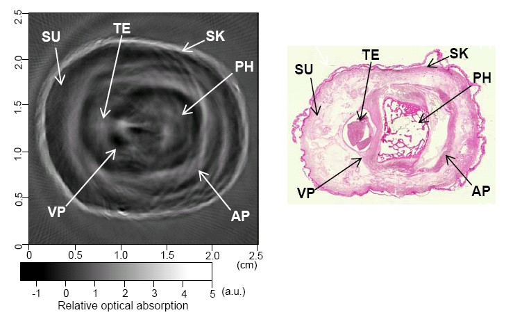
(Left) Cross-section photoacoustic image of a human finger joint. (Right) Histological photograph of the imaged cross section (gold standard).
AP: aponeurosis; PH: phalanx; SK: skin; SU: subcutaneous tissue; TE: tendon; VP: volar plate.
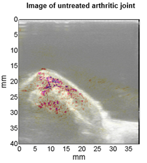
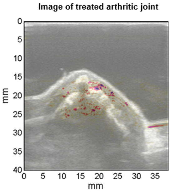
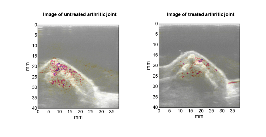
Photoacoustic image (pseudo-color) superimposed on the gray scale ultrasound image of rat ankle joints. (Left) An arthritic joint before treatment. (Right) An arthritic joint after treatment. The difference between the two images suggests the capability of photoacoustic imaging in grading synovitis, and in characterizing the functional change in response to treatment.
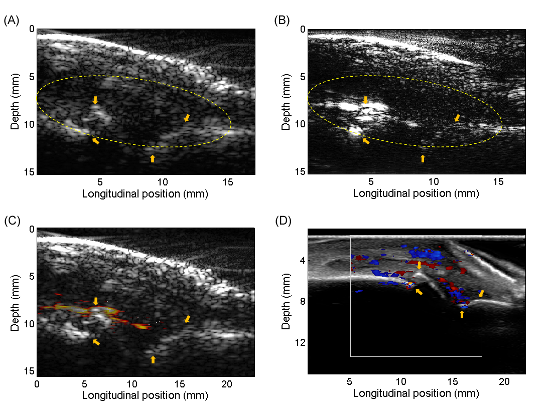
Photoacoustic (PA) and ultrasound (US) dual imaging of a metacarpophalangeal (MCP) joint of a patient affected by inflammatory arthritis. (A) Gray-scale B-mode US image. (B) Gray-scale PA image of the same imaging plane. (C) Pseudocolor PA image super-imposed on the gray scale US image, demonstrating the active vascularity in the joint. (D) Gold-standard US Doppler image acquired by a commercial US unit confirming the active synovitis in the studied joint.