
Introduction:
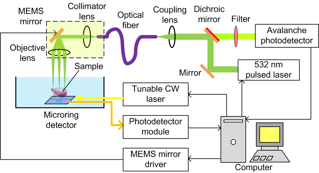
A part of our research is focused on the development and application of photoacoustic microscopy (PAM) which offers image quality similar to conventional optical microscopic techniques but better imaging depth and unique optical absorption contrast. By collaborating with Prof. Jay Guo at U-M EECS, we have developed an “all optical” PAM system. Instead of using conventional PZT transducers, this PAM system employs an optical microring resonator as the ultrasonic detector. Due to the high sensitivity and drastically increased bandwidth for ultrasonic detection facilitated by the microring, our PAM system allows excellent depth sectioning and has achieved an axial resolution better than 5 micrometer. The all-optical PAM system has been further developed and miniaturized to satisfy the requirements of clinical endomicroscopy. It is a consensus that endoscopy could one of the most promising application of the emerging biomedical photoacoustic technology. In our lab at Michigan, photoacoustic microscopy is adapted to transurethral imaging of bladder cancer and to transrectal imaging of inflammatory bowel disease.

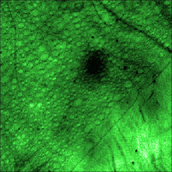
(A) Schematic of a photoacoustic microscopy (PAM) and confocal fluorescent microscopy (CFM) dual-modality imaging system. (B) An example PAM image showing the microvasculature in a biological sample. (C) An example CFM image showing the cellular distribution in a biological sample.
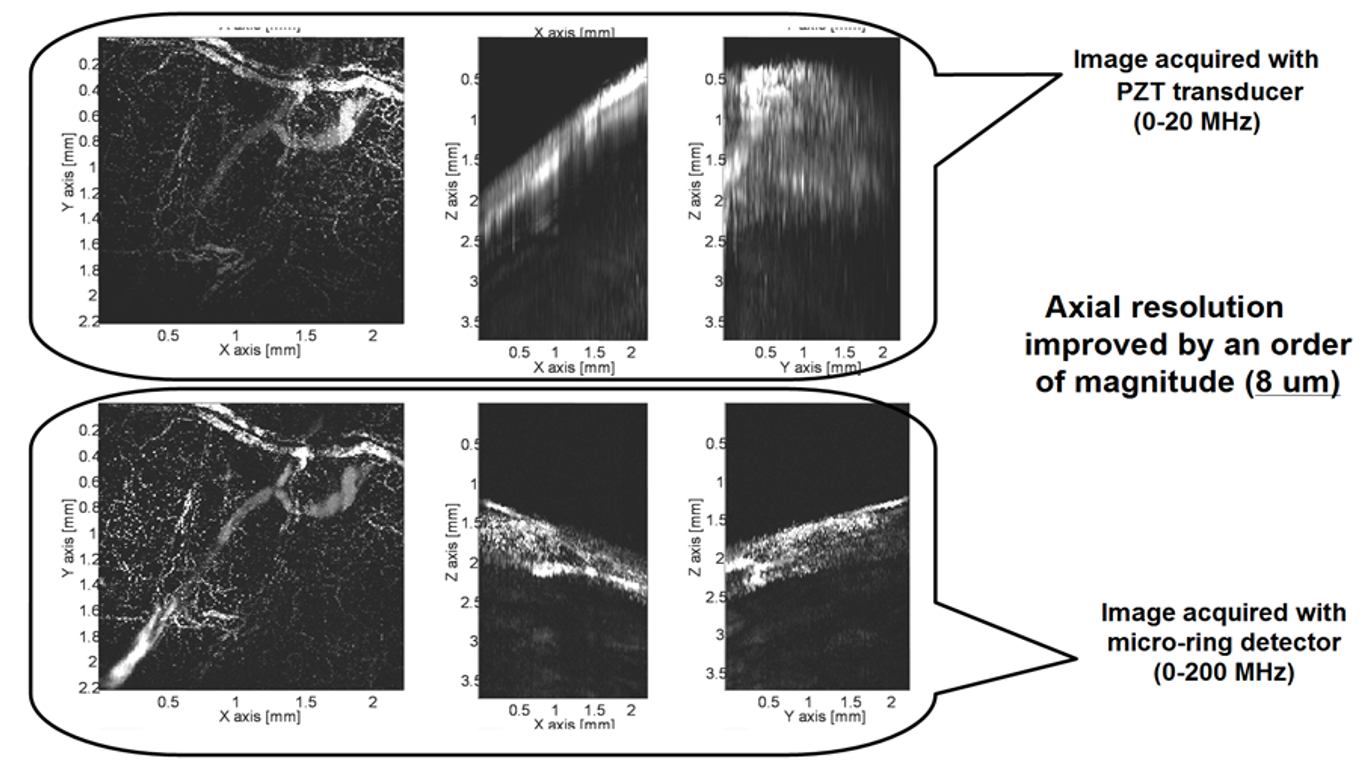
(Upper) In vivo photoacoustic microscopic image of the vasculature in a mouse ear acquired with a conventional PZT transducer. (Lower) The image from the same sample acquired with a super-broad bandwidth micro-ring detector. Significant improvement in axial resolution facilitated by the micro-ring detector can be noticed.
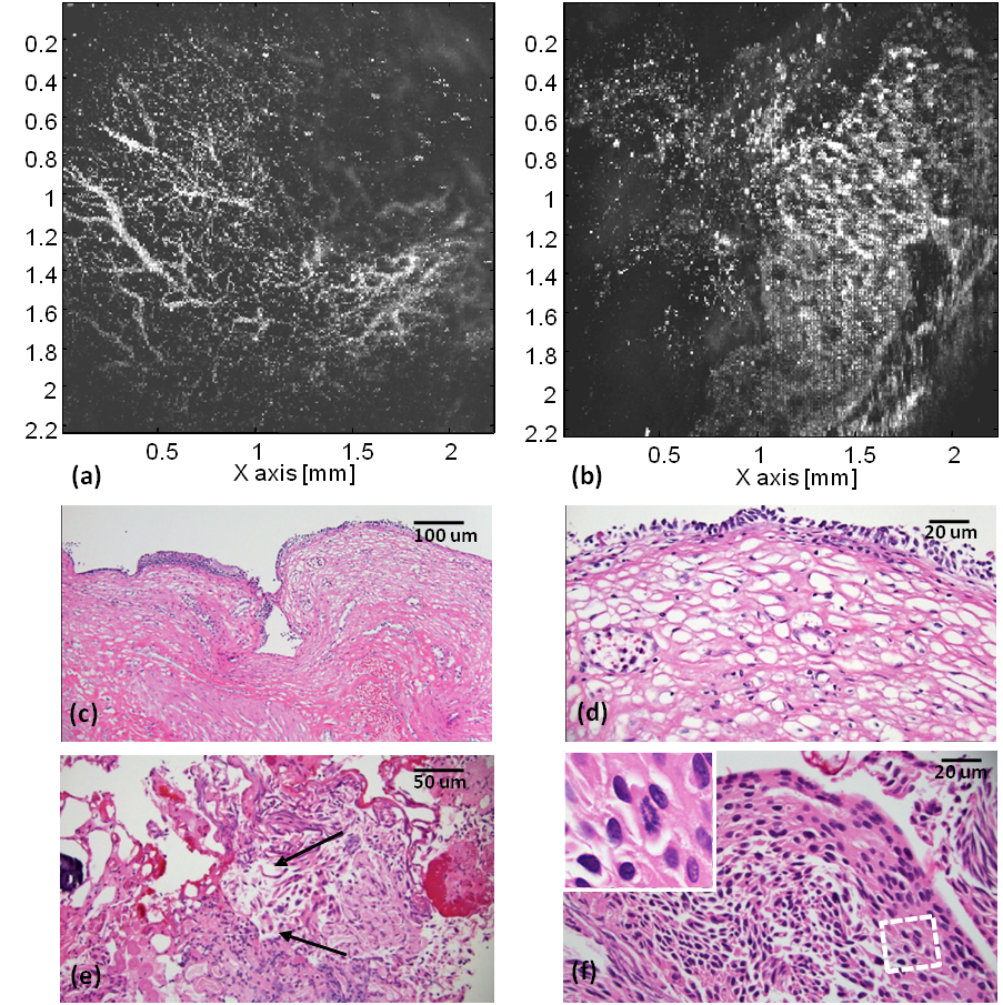
Photoacoustic microscopy (PAM) and histological assay of normal and cancerous human bladder specimens. PAM images of (a) a normal and (b) a tumor specimen. (c) Low magnification and (d) high magnification histological photographs of the normal specimen sectioned down from the area of (a). (e) High magnification histological photograph of the tumor specimen depicting infiltration of large disordered cells into superficial lamina propria (marked by arrows). (f) Another histological photograph of the tumor depicting the loss of polarization and anisocytosis. The inset is the magnification of the marked area presenting the rare mitotic figures.