
Introduction:
Optical signal is highly sensitive to the molecular conformation of biological tissues which can aid in describing tissue physiological and biochemical changes. Intrinsically, the dominant chromophores in the breast are oxy- and deoxy-hemoglobin in the near-infrared (NIR) region. Hence, optical imaging modalities measuring optical absorption are unique in mapping and quantifying vascularization and hemoglobin oxygen saturation of biological tissues. However, due to the overwhelming scattering of light in biological tissues, conventional optical imaging has very limited spatial resolution in visualizing tumors in deep breast tissues. Moreover, the diagnostic information extracted from conventional optical imaging results is very difficult to be quantified. Photoacoustic tomography (PAT) is an emerging hybrid technology combining the merits of light and ultrasound to achieve both high optical contrast and good spatial resolution in imaging tissue structures and functions upon progression and regression of cancer. With the unique features, PAT, as a natural and promising complement to existing US technologies, could contribute to the detection, diagnosis and prognosis of breast cancer.
Example results:
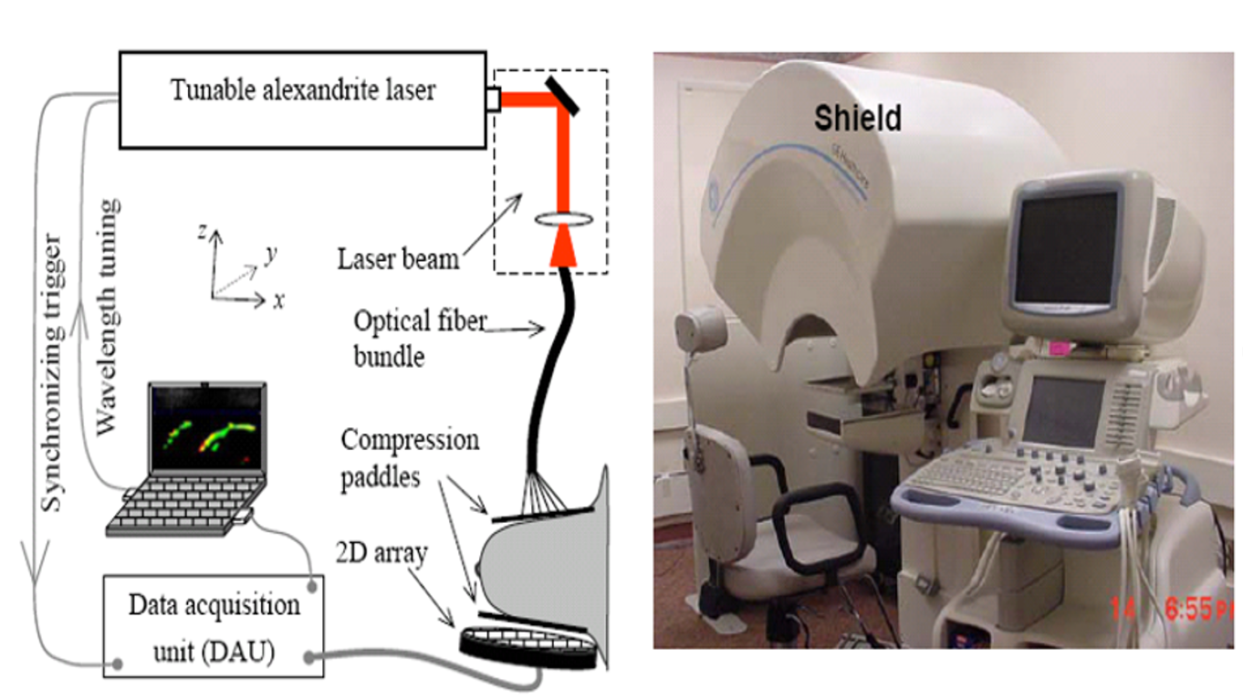
(Left) Photoacoustic tomography (PAT) of breast cancer in a mammographic geometry. (Right) A platform combining three medical imaging modalities for breast cancer detection and diagnosis: 3D x-ray tomo, automatic ultrasound (US), and PAT.
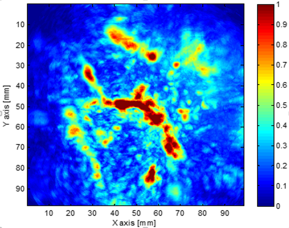
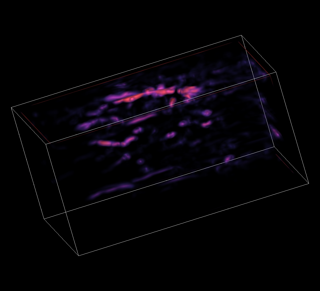
In vivo photoacoustic image of vasculature in a normal breast. (Left) 2D maximum amplitude projection image. (Right) 3D image of the vasculature.